
Diagram Of Hip Muscles And Tendons : Adductor Muscles Of The Hip Tendon Anatomy Muscles Of The Hip Text Hand Human Png Pngwing - A tendon is the end part of a muscle that attaches the muscle to the bone.. • to explain how movement is brought. In human anatomy, the muscles of the hip joint are those muscles that cause movement in the hip. Tensor faschia latae is the muscle that controls what? Physical therapy can improve joint mobility, range of motion, and muscle strength. Each of these muscles is a discrete organ constructed of skeletal muscle tissue, blood vessels, tendons, and nerves.
Hip joint ligaments and tendons, lower back muscle pain. Learn how they work together. The anterior muscles of the hip allow for rotational movements and flexion of the hip as well as flexion of the vertebral column, but only when they apply their contraction during cohesive unison. The muscle that is contracting is called the agonist and the muscle that is relaxing or lengthening is called the antagonist. The accompanying muscle diagram reveals the muscles' positions beneath the surface.
Each of these muscles is a discrete organ constructed of skeletal muscle tissue, blood vessels, tendons, and nerves.
General causes of hip pain include The core muscles are those in the abdomen, back, and pelvis, and they also stabilize the body and assist in tasks, such as lifting weights. The two heads unite and spread into an aponeurosis which is prolonged downward on the anterior surface of the muscle, and from this, the muscular fibers arise. Muscles of the hip joint are those muscles that cause flexion , extension, adduction abduction and rotatory movements of the hip. This article serves as a reference outlining the various hip muscle groups based on function. Flexion of hip and vertebral column. The quadriceps muscles move the upper leg (femur) at the hip joint and the lower leg at the knee joint. The iliopsoas not only functions as the prime mover of hip flexion, it also plays an essential role in the functional stability of the hip, the pelvis, and even the spine (fig. Physical therapy can improve joint mobility, range of motion, and muscle strength. Sartorius is a unique muscle because it is the only knee flexor that originates anteriorly. The magnitude and timing of mtu length peaks were each compared between walking and running. The muscle that is contracting is called the agonist and the muscle that is relaxing or lengthening is called the antagonist. Nine may seem like quite a lot, but these muscles are essential for creating the wide range of hip movements used by dancers, sportspeople and music lovers.
The two heads unite and spread into an aponeurosis which is prolonged downward on the anterior surface of the muscle, and from this, the muscular fibers arise. Ligaments are soft tissue structures that connect bones to bones. Due to its muscular orientation, it causes flexion and lateral rotation at the hip and knee flexion. Hip pain areas and causes hip skeleton hip and spine diagram hip illustration posterior hip bone anatomy leg hip muscle anatomy hip and femur anatomy anterior hip ligaments left hip bone anatomy hip s3.amazonaws.com. In addition, weakness of the buttock muscles and hip rotators generally occurs because of the loss of movement.

Muscle and tendons in a diagram of the arm;
The psoas major, the psoas minor, and the iliacus (fig. The ligaments, tendons, and muscles in the hip joint play a vital role in your ability to walk, run, move, and exercise. Muscles and tendons of the hand. While this density makes the tendons stronger, the lack of elasticity of the tendon and the constant pulling on its attachment to the bone with movement, makes it other areas where tendonitis occurs include the hips and ankles. The anterior muscles of the hip allow for rotational movements and flexion of the hip as well as flexion of the vertebral column, but only when they apply their contraction during cohesive unison. In addition, weakness of the buttock muscles and hip rotators generally occurs because of the loss of movement. • to state the function of cartilage &. In human anatomy, the muscles of the hip joint are those muscles that cause movement in the hip. Muscle and tendons in a diagram of the arm; The hip muscles are all the muscles that act on the hip joint. Diagram showing the changes that occur in tendons from inflammatory tenosynovitis through. Muscles/tendons flashcards from molly m. The core muscles are those in the abdomen, back, and pelvis, and they also stabilize the body and assist in tasks, such as lifting weights.
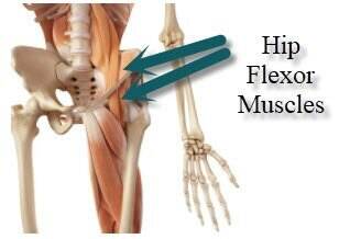
While this density makes the tendons stronger, the lack of elasticity of the tendon and the constant pulling on its attachment to the bone with movement, makes it other areas where tendonitis occurs include the hips and ankles. Each of these muscles is a discrete organ constructed of skeletal muscle tissue, blood vessels, tendons, and nerves. This diagram with labels depicts and explains the details of hip muscles and tendons. Sartorius is a unique muscle because it is the only knee flexor that originates anteriorly. The core muscles are those in the abdomen, back, and pelvis, and they also stabilize the body and assist in tasks, such as lifting weights.
Ligaments, tendons, and muscles play an important role in the function of the hip.
Learn vocabulary, terms and more with flashcards, games and other study tools. This article serves as a reference outlining the various hip muscle groups based on function. Distribution on the hip can be shown. Related online courses on physioplus. • coils and patient position: The anterior muscles of the hip allow for rotational movements and flexion of the hip as well as flexion of the vertebral column, but only when they apply their contraction during cohesive unison. Tight muscles, tendons, ligaments, and tissues occur with osteoarthritis further limiting joint movement. Tensor faschia latae is the muscle that controls what? In an antagonistic muscle pair as one muscle contracts the other muscle relaxes or lengthens. Muscle and tendons in a diagram of the arm; A tendon is the end part of a muscle that attaches the muscle to the bone. A thickened area of tendon known as the iliotibital tract serves as a secondary insertion point, which is. The core muscles are those in the abdomen, back, and pelvis, and they also stabilize the body and assist in tasks, such as lifting weights.
Muscle and tendons in a diagram of the arm; hip muscles diagram. Hip joint ligaments and tendons, lower back muscle pain.